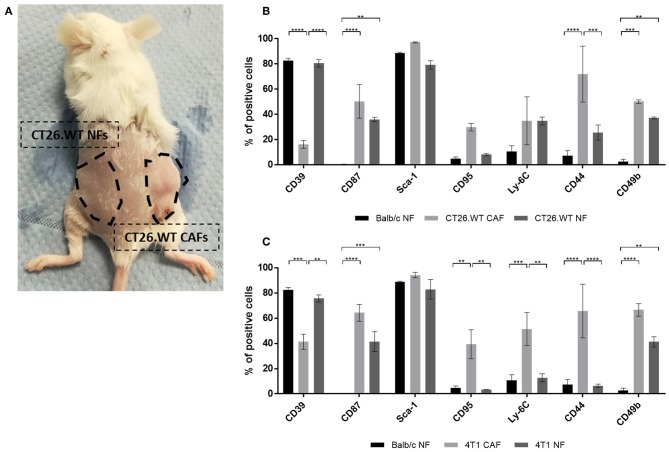
Figure 5.
Ex vivo analysis of normal fibroblasts from tumor-bearing mice. (A–C) Comparison of NFs derived from tumor-bearing mice with “healthy” NFs and CAFs. Fibroblasts were isolated and analyzed ex vivo for their expression of CD39, CD87, Sca-1, CD95, Ly-6C, CD44, and CD49b. NFs from tumor-bearing mice were isolated from tumor-free skin and mammary fat pad of the flank where no tumor cells had been injected (A), thus they have not been in direct contact with tumor cells. As healthy NF control population, fibroblasts were isolated from healthy BALB/c mice. The phenotype of NFs from tumor bearing mice was analyzed in CT26.WT (B) and 4T1 (C) mouse tumor model. The percentage of cells expressing the respective marker is depicted. Data are reported as median and SEMs for three replicates from at least two independent experiments and statistical analysis was performed using the two-way ANOVA (**P < 0.005; ***P < 0.0005; ****P < 0.0001).

