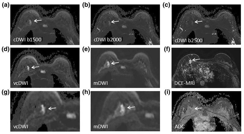
FIGURE 1:
A 34-year-old woman with DCIS in left breast. cDWI (A–C), vcDWI (D,G), mDWI with b-value of 1000 s/mm2 (E,H), DCEMRI (F), and ADC (I) are shown. G,H are magnified views. As b-value increases, the signal intensity of background fibroglandular tissue decreases, as well as that of cancer tissue. The signal intensity of cancer on vcDWI is preserved well with background suppression, and the lesion is more conspicuous and sharper on vcDWI than on cDWIs and mDWI, which allows the lesion to be more easily detected and characterized. The diagnostic BI-RADS scores given by Reader X.H.Z. and Reader W.M.H. were 5, 5, respectively, which correctly diagnose the lesion as malignant.

