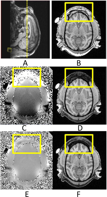
Fig 3. Structural images of an adult macaque head. A: Sagittal T1-weighted structural Image of the monkey head. B: Axial T1-weighted image without fat suppression;
C: The original field map; D: T1-weighted image with fat suppression; E: The field map after re-shimming with GRESHIM; D: T1-weigted image with fat suppression.
The square boxes show the interested area with susceptibility effects.

