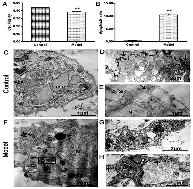
Fig 1.
(A) The cell viability in the model group was significantly lower compared with the control group. (B) The apoptosis cells were much higher in model group than those in the control group (n = 3 per group, **P < 0.01: the control group vs. the model group). (C) The normal chondrocytes had abundant mitochondria and rough endoplasmic reticulum. The enlargement version showed that lanthanum ions precipitated outside the cell membrane (arrows, D and E). (F) The model group showed that IL-1β-induced chondrocytes had vacuolization and mitochondrial pyknosis, the tracer lanthanum ions appeared in the organelles or lysosomes. The enlargement version showed that lanthanum ions scattered in organelle and cytoplasm (arrows, G and H). (N) Nucleus, (Mi) mitochondria, (RER) rough endoplasmic reticulum, (Ly) lysosome, (G) lanthanum ions (arrows), mitochondria dissolved (rectangle), vacuolized endoplasmic reticulum (△).

