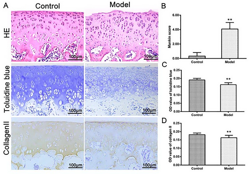
Fig 4.
(A) HE stained showed a complete structure in normal cartilage, however, the model group showed the cartilage surface damaged with chondrocytes proliferated; Toluidine blue staining and collagen II showed a modest decrease in the model group compared with those in the control group, as indicated by the semi-quantitative analysis B, C and D (n = 12 per group, **P < 0.01: the control group vs. the model group).

