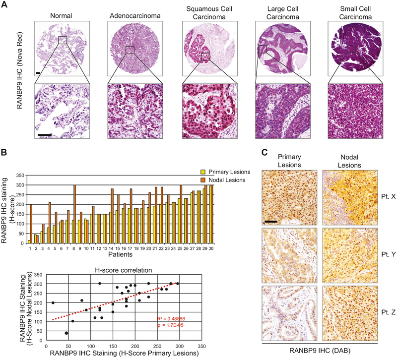
Fig. 1.
RANBP9 is overexpressed in lung cancer primary tumors and nodal metastases. a Representative examples of RANBP9 expression evaluated by IHC (Nova Red staining) in different histotypes of lung cancer and in normal adjacent tissue (NAT). Scale bars represent 100 μm. b Upper panel: RANBP9 protein levels evaluated by IHC in 30 cases of NSCLC primary tumors (yellow bars) and corresponding nodal metastasis (orange bars). Lower panel: positive correlation of RANBP9 protein levels between the primary lung adenocarcinomas and the nodal lesions (R2 = 0.48886; p < 0.001). The correlation was calculated according to Pearson correlation coefficient. Trend is indicated by a red dotted line. c Representative examples of RANBP9 expression in primary tumors and in the corresponding nodal lesions evaluated by IHC (DAB staining) in three different, representative patient specimens. Scale bars represent 100 μm

