Fig. 3.
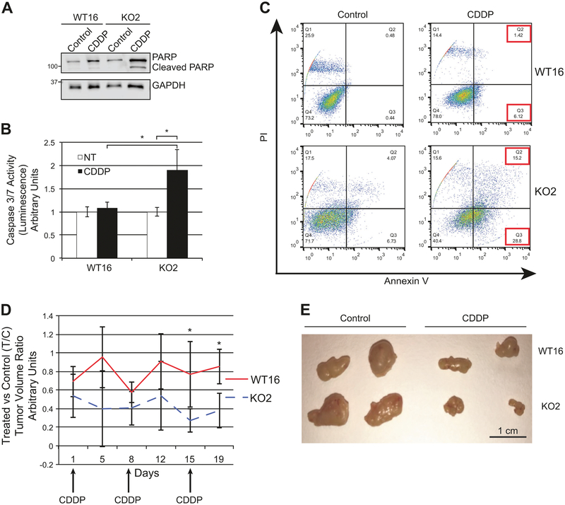
RANBP9 KO are more sensitive to cisplatin than RANBP9 WT NSCLC cells both in vitro and in vivo. a Western blot analysis of RANBP9 WT and KO A549 clones, treated for 24 h with 15 μM CDDP to evaluate total and cleaved PARP. GAPDH was used as loading control. b Caspase 3/7 activation assay performed on A549 RANBP9 WT and KO clones. Cells were left untreated or treated with CDDP 15 μM for 24 h. *p < 0.05. Data are representative of three independent experiments performed in triplicate. c Flow cytometry analysis of RANBP9 WT and KO cells left untreated or treated with CDDP 20 μM for 72 h, and stained with propidium iodide (PI) and Annexin V-FITC. Data are representative of two independent experiments. d NOD-SCID mice (n = 8) were subcutaneously injected with 5 × 10⁶ A549 RANBP9 WT and KO on the left and right flank, respectively. Once the tumors were detectable, mice were treated with control solution (n = 4) or 5 mg/kg of CDDP (n = 4), once a week for three weeks. Mice were monitored and tumors were measured by physical examination twice a week. After 19 days from the first injection, mice were euthanized. Tumors were excised and measured. CDDP-treated versus control-treated tumor volume ratios (T/C) ± S.D. are reported. *p-value < 0.05. e Representative image of RANBP9 WT and KO tumors, treated with CDDP and vehicle control treated

