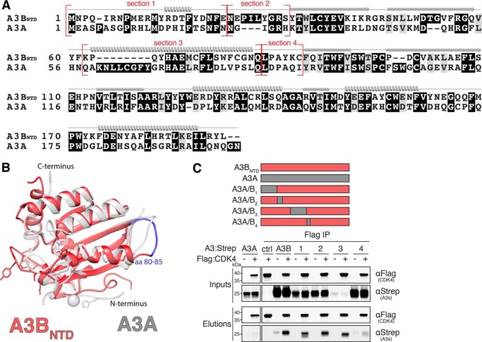
Figure 4.
CDK4 interacts with a N-terminal region of A3B. A, amino acid alignment of A3BNTD and A3A with chimeric junctions indicated in red and structural elements shown above the alignment (α helices, β strands, and loop regions). B, ribbon schematic overlay of the crystal structures of A3BNTD (red, PDB 5TKM) and A3A (gray, PDB 4XXO). A3BNTD residues 80–85 are highlighted in blue (see text for details). C, anti-Flag (CDK4) co-IP of indicated Strep-tagged A3BNTD constructs from 293T cells. Parallel reactions were done with empty vector and A3A-S as negative controls. See Fig. 2A legend for a description of the IP labeling scheme.

