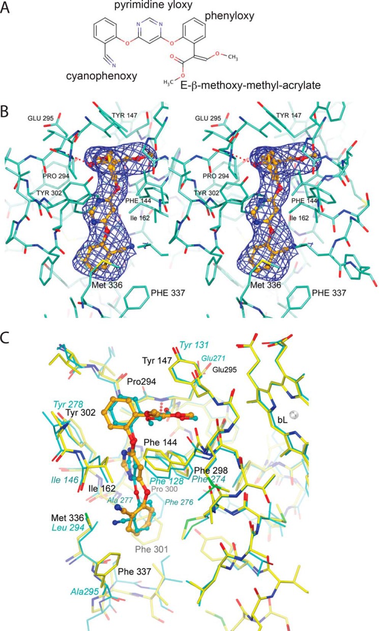
Figure 4.
Chemical structure of azoxystrobin and its binding environment in Rsbc1. A, chemical structure of azoxystrobin. B, stereographic diagram of the bound azoxystrobin in stick model in the QP pocket overlaid with the blue difference electron density cage for azoxystrobin. The electron density was generated with phenix.polder and contoured at 5σ. Residues lining the QP pocket are shown as a stick model. The hydrogen bond between azoxystrobin and the main-chain amide N atom of Glu-295 is shown as the dotted line in red. C, superposition of the QP site between the Btbc1/azo and Rsbc1/azo structures. The poses of azoxystrobin molecules bound to the QP sites of Rsbc1 (yellow sticks) or Btbc1 (cyan sticks) are nearly identical.

