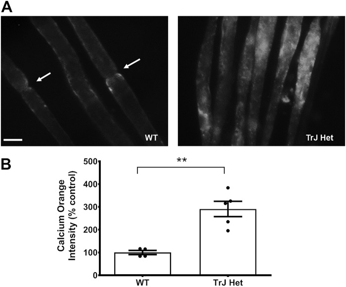
Figure 6.
Intracellular calcium levels are increased in TrJ nerves. A, sciatic nerves from 4- to 8-week-old WT and TrJ-Het mice. The perineurium was extracted, and fibers were teased apart before being incubated with Calcium Orange AM in DMEM. Nerves were visualized at ×400 on a fluorescent microscope. Scale bar, 5 μm. B, average signal intensities were obtained and normalized to the area of the regions of interest and the mean intensity of the neighboring background. The nodal regions in the WT nerves (arrows) were avoided because they exhibited higher levels of Ca2+ than the internodal regions. n = 3, **, p = 0.0018, based on a Student's t test.

