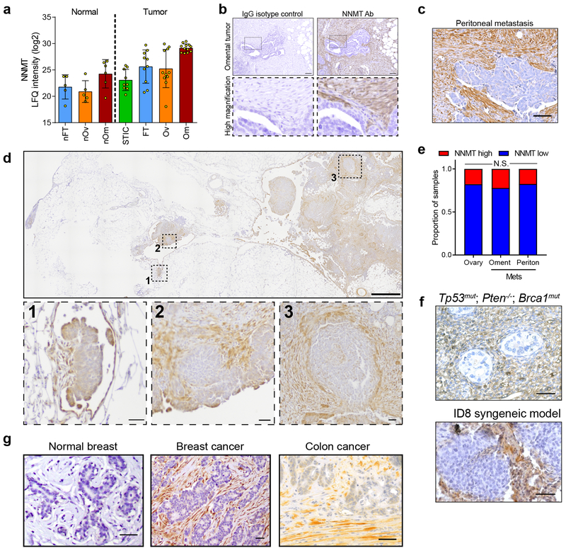
Extended Data Fig. 3: NNMT is highly expressed in the stroma of ovarian cancers.
(a) Quantitative proteomics of the stroma finds elevated expression of NNMT in omental metastases (n = 11) compared to normal (normal fallopian tube (nFT; n = 5), normal ovarian (nOv; n = 5), and normal omentum (nOm; n = 6) and primary tumor tissues (STIC, n = 9; invasive fallopian tube (FT), n = 10; and invasive ovarian (Ov), n = 11). (b) Human omental metastasis tissue stained with NNMT-specific antibody or IgG isotype control; no non-specific staining is observed. Scale bar = 50 μm. (c) Representative NNMT IHC of peritoneal metastasis. (d) NNMT IHC of an omentum with micrometastases (1) and larger metastases (2 and 3). NNMT is detected in the stroma of very early metastases. (e) Quantification of tumor NNMT staining in TMA analysis. Chi-square test, n = 169 ovarian, 135 omental, and 92 peritoneal samples. (f) Representative IHC of NNMT in the stroma of metastases in an autochthonous model of ovarian cancer (top, PAX8:TP53mut;PTEN−/− ;BRCA1mut) and a syngeneic model (bottom, ID8 intraperitoneal xenograft). (g) NNMT is expressed in the stroma of breast and colon cancers, but not normal breast stroma.

