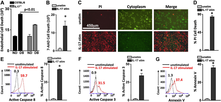
Figure 3. IL-17A elicits apoptosis in human retinal endothelial cells.
Quantification of flow cytometry analysis (n=6) of retinal endothelial cell death (CD144+/7-AAD+) 18h after murine retinal endothelial cells were co-cultured with bone marrow cells from non-diabetic (ND) and diabetic (DB) C57BL/6 (black) or IL17A−/− (grey) mice (A), or of human retinal endothelial cells stimulated with recombinant human IL-17A (B). C) Representative microscopy of unstimulated (top) and IL-17A stimulated (bottom) human retinal endothelial cells (hREC) 48h after incubation; dead red cells are Propidium iodide (PI) positive and viable green cells retain the FITC cytoplasmic membrane stain (scale bars of all images = 450μm). D) PI quantification of flow cytometry analysis of 6 separate unstimulated (grey) and IL-17 stimulated (black) hREC samples. Representative flow cytometry analysis of unstimulated (black) and IL-17A stimulated (red) hRECs, as well as the quantified percent of apoptotic cells using Active Caspase 8 (E), Active Caspase 3 (F), and Annexin V assays (G) 6h after stimulation. Numbers above overlay peaks represent percent positive cells of 30,000 events, and the graph displays the percentage of Caspase 8 or 3, and Annexin V positive cells in six separate samples. *=p<0.01 per unpaired student’s t-test. Data are representative of three separate experiments with similar results.

