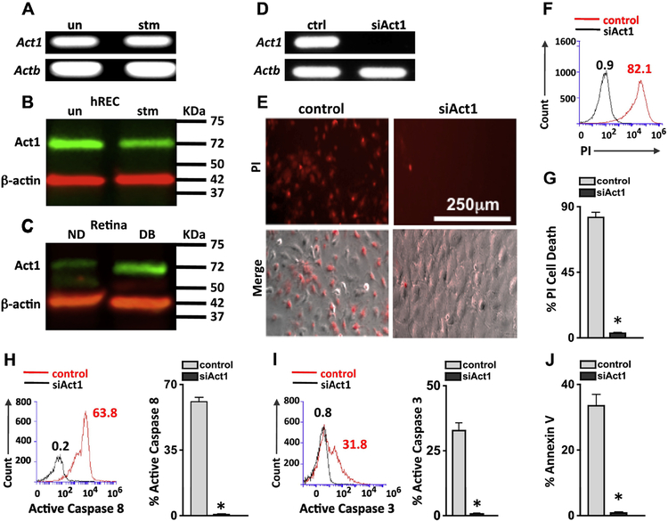
Figure 4. Act1 dependent retinal endothelial cell death.
A) Qualitative qPCR analysis of Act1 expression in mRNA of unstimulated and IL-17A stimulated hREC. Actb was used as a loading control. Immunoblot analysis of Act1 in protein lysates of unstimulated (un) or IL-17A stimulated (stm) hREC cultures (B), and non-diabetic (ND) and diabetic (DB) C57BL/6 mouse retinas (C); protein was collected 2-months post-diabetes or 6h post-stimulation. β-actin was used as a loading control. D) Act1 analysis by RT-PCR verifying Act1 gene silencing (siAct1) in hREC cultures; ctrl samples were transfected with scrambled sequences. E) Representative images of IL-17A stimulated control and siAct1 hREC viability; 48h after stimulation. Dead cells were examined using Propidium Iodide (PI) and Phase (Merge (PI + Phase)) microscopy analysis (scale bars for all images = 250 μm). Representative overlay of PI flow cytometry analysis in scrambled control (red) and siAct1 (black) hREC (F), and percentage of scramble control (grey) and siAct1 (black) PI+ cells (G). Representative flow cytometry analysis (left) and quantification (right) of 6 separate samples of active Caspase 8 (H) and active Caspase 3 (I) in IL-17A stimulated control and siAct1 hREC cultures 6h after incubation. Numbers represent percent positive cells of 30,000 events. J) Percentage of apoptotic Annexin V positive cells per flow cytometry analysis of 6 samples/group. *=p<0.01 per unpaired student’s t-test. Data are representative of two separate experiments with similar results.

