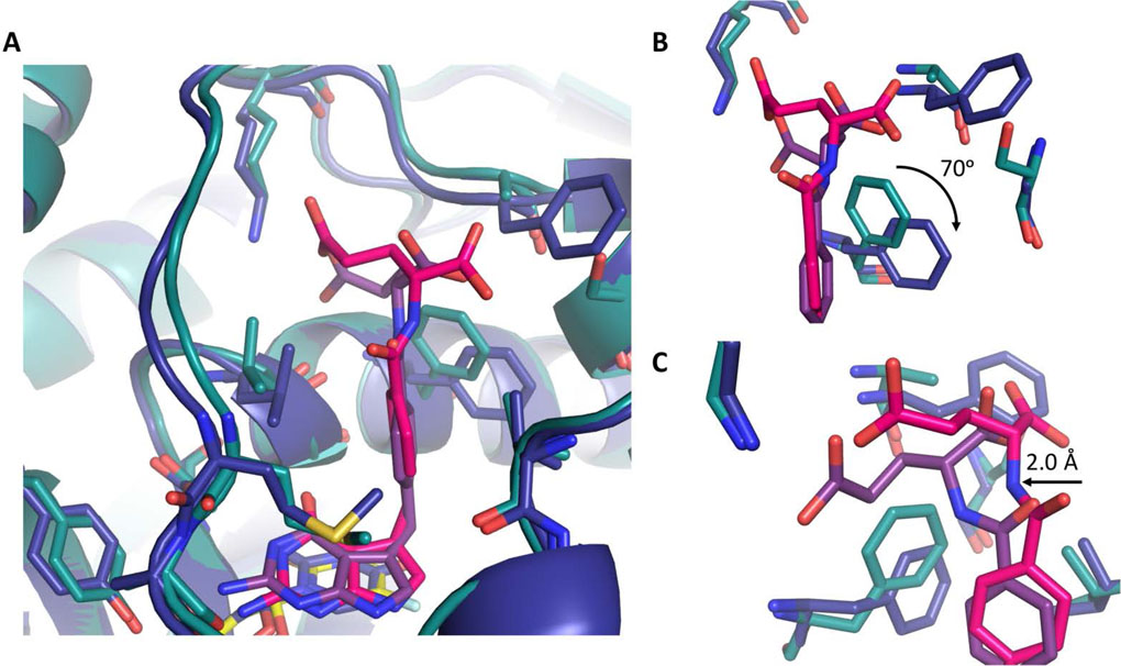
Figure:4.
Comparison of ChTS (green) bound to 2 (magenta) and FdUMP (yellow) with hTS (blue) bound to 2 (purple) and dUMP (yellow). A. Comparison of 2 bound in ChTS and hTS active site. B. Zoom in view hTS residue F225 position versus ChTS residue F433 upon binding of 2. C. Zoom in view of glutamate moiety of 2 interactions with both ChTS and hTS. Rotation of hTS residue F225 provides greater access allowing 2 to shift deeper into active site.

