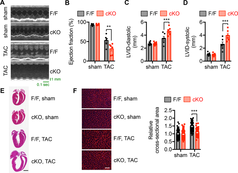
Figure 2. XBP1s deficiency exacerbates heart failure progression by pressure overload.
A. Control and cKO animals of 8 weeks old were used for sham or TAC surgery. Cardiac function was determined 2 weeks later. Representative echocardiography images of control and cKO animals after sham or TAC surgery are shown.
B. XBP1s deficiency in the heart led to deterioration of cardiac function and accelerated progression to cardiomyopathy, as revealed by a decrease in ejection fraction (%). N = 6–9.
C. Diastolic LVID was significantly elevated in the cKO mice after TAC. N = 6–13.
D. LVID at systole was increased by XBP1s deficiency in the heart. N = 6–13.
E. Representative cardiac images showed deficiency of ventricular growth in the cKO mice after TAC. Scale bar: 2 mm.
F. Wheat germ agglutinin (WGA) staining was performed to visualize cardiac myocytes (left). Scale bar: 50 μm. Quantification is shown at the right. N = 209 for sham/FF; 188 for sham/cKO; 115 for TAC/FF; 143 for TAC/cKO. Two-way ANOVA analysis was conducted, followed by Tukey’s test. **, P<0.01; ***, P<0.001.

