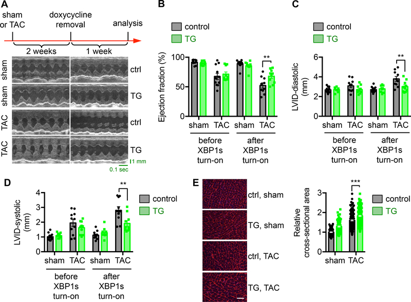
Figure 3. Overexpression of XBP1s in the heart improves cardiac function in response to pressure overload.
A. XBP1s expression was turned on after 2 weeks of TAC, before the onset of cardiac dysfunction (indicated as doxycycline removal). After another week, control animals (single TRE-XBP1s or αMHC-tTA transgenics alone under water without doxycycline) displayed cardiomyopathy and cardiac dysfunction, while transgenic mice (TRE-XBP1s and αMHC-tTA double transgenics under water without doxycycline) showed significant improvements in cardiac function, as evidenced by representative echocardiographic images.
B. Ejection fraction (%) was significantly increased by XBP1s overexpression. N = 9–13.
C. LVID at diastole was reduced in the XBP1s transgenic mice after TAC. N = 9–13.
D. Systolic LVID in the transgenic mice was improved. N = 9–13.
E. WGA staining was conducted (left). Scale bar: 50 μm. Quantification of the relative cross-sectional area shows significant upregulation of cardiomyocyte size by XBP1s overexpression (right). N = 128 for sham/control; 104 for sham/TG; 198 for TAC/control; 226 for TAC/TG. Two-way ANOVA analysis was conducted, followed by Tukey’s test. **, P<0.01; ***, P<0.001.

