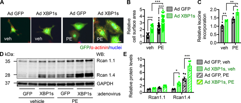
Figure 5. XBP1s expression is sufficient to stimulate cardiomyocyte hypertrophic growth.
A. XBP1s overexpression in cardiomyocytes led to an increase in cell size. NRVMs were infected by adenovirus expressing control GFP or XBP1s, followed by PE treatment. Note that the XBP1s-expressing virus is bi-cistronic for GFP and XBP1s, and GFP positivity indicates infection by either GFP-only control or XBP1s-GFP virus. Scale bar: 20 μm.
B. Quantification of A. N = 34 for veh/Ad GFP; 33 for veh/Ad XBP1s; 51 for PE/Ad GFP; 107 for PE/Ad XBP1s.
C. Protein synthesis was elevated by XBP1s overexpression as revealed by 3H-leucine incorporation. N = 3 for PE/Ad XBP1s. N = 6 for the other groups.
D. XBP1s overexpression potentiated PE-induced upregulation of Rcan1.4 at the protein level. As a control, Rcan1.1 did not show a similar trend.
E. Quantification of D. N = 5 for Rcan1.4/Ad XBP1s. N = 6 for the other groups. Two-way ANOVA analysis was conducted, followed by Tukey’s test. *, P<0.05; **, P<0.01; ***, P<0.001.

