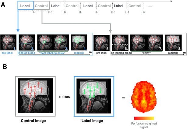
Figure 1.
How ASL works. (A) Sequence of events occurring during ASL image acquisition. During labeling (blue), the following steps occur. 1. As the subject is placed in the MRI scanner and in the absence of any MR pulses, the body's protons, including those in blood-water (red), are aligned with the main magnetic field. 2. Blood–water (red) flowing through the labeling region (in this case a plane; blue dotted line), is rotated out of phase with the main magnetic field. Labeled blood is displayed in green. 3. The “postlabeling delay” is equivalent to a “waiting period” wherein labeled blood (green) is able to flow from the labeling region to brain tissue before the images are collected. 4. Images are acquired that capture the magnitude of labeled blood (green) that has arrived at the tissue. During the acquisition of control images (gray), the same sequence of steps occurs except no blood is labeled. 5. Blood–water (red) is aligned with the main magnetic field. 6. Blood–water (red) flowing through the labeling region (dotted line) are NOT rotated out of phase with the main magnetic field. This means that the blood water is not magnetically labeled. 7. A waiting period identical to the “postlabeling delay” is allowed to elapse, except that here the blood is not labeled (red). 8. Images are acquired that capture the magnitude of background “static” tissue signal where no blood is labeled. (B) During postprocessing, the subtraction between “control” and “label” volumes allows for the calculation of perfusion-weighted maps. The later can be further processed to obtain an rCBF map that is fully quantitative (mL blood/100 g tissue/min). ASL, arterial spin labeling; rCBF, regional cerebral blood flow; TR, repetition time.

