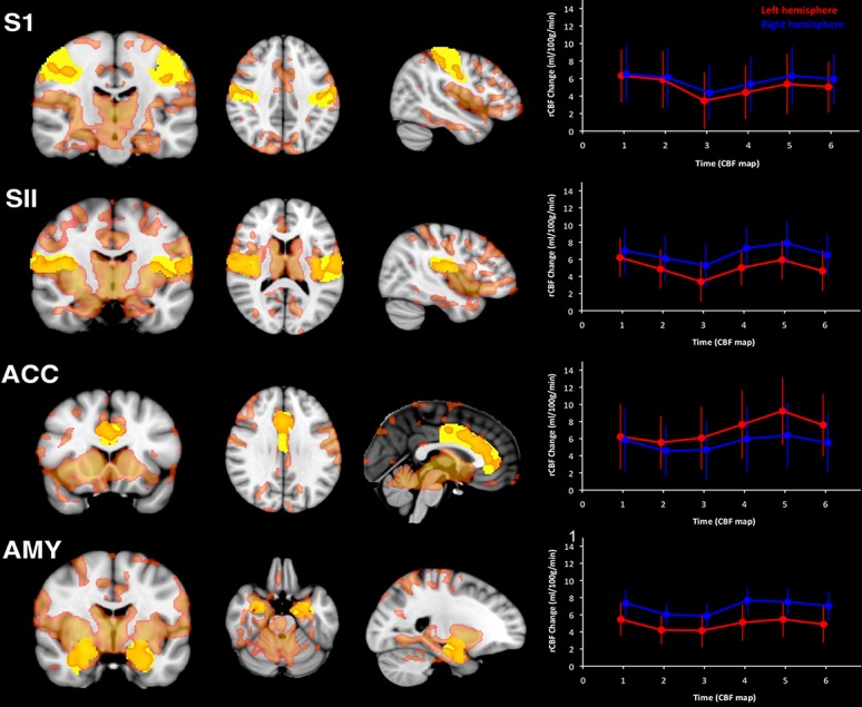
Figure 5.

Imaging postsurgical pain with ASL. Elevations in rCBF immediately after third molar tooth extraction. Voxels in red designate significant increases in rCBF postsurgery. A priori defined regions of interest (ROIs) for key anatomical areas of the brain are in yellow. Line plots indicate the magnitude of the increase in rCBF during postsurgical pain, compared with pain-free presurgical periods in each ROI in left hemisphere (red line) and right hemisphere (blue line). Each data point represents the mean rCBF increase (in millilitres/100 g/min) during each individual pCASL scan. ACC, anterior cingulate cortex; AMY, amygdala; S1, primary somatosensory cortex; SII, secondary somatosensory cortex (from Ref. 34). ASL, arterial spin labeling; pCASL, pseudocontinuous ASL; rCBF, regional cerebral blood flow.
