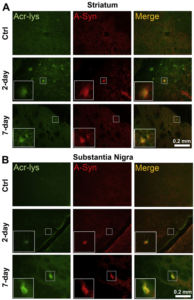
Figure 11.
Post-injury elevation of co-localization of FDP-lysine and α-syn immunoreactivities. A) Representative immunofluorescence staining in the striatum shows sham Control (“Ctrl”) 2-day and 7-day post injury. Acrolein shown in green and α-syn in red, with areas of co-localization (“Merged”) appearing yellow. (B) Corresponding images for the substantia nigra region. White zoom boxes represent areas of increased immunoreactive staining intensity.

