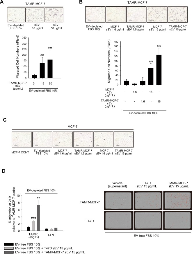
Figure 7.
Essential role of sEV in cell migration of TAMR-MCF-7 cells. (A) Migration of TAMR-MCF-7 cells after incubation with TAMR-MCF-7 sEV (16 and 50 μg/mL) for 18 h (n = 10). (B) Migration of TAMR-MCF-7 cells after incubation with MCF-7 or TAMR-MCF-7 sEV (1.6 and 16 μg/mL) for 18 (n = 10). (C) Migration of MCF-7 cells after incubation with MCF-7 or TAMR-MCF-7 sEV (1.6 and 16 μg/mL) for 18 h (n = 10). Cell migration was evaluated by Transwell migration assay in sEV-depleted FBS condition. (D) Migration of T47D and TAMR-MCF-7 cells after incubation with T47D and TAMR-MCF-7 sEV (15 μg/mL) for 24 h (n = 3). Migrated cells were marked in red circle by image analysis. Images were taken by Incucyte Zoom. All data represent the mean ± SE (###p < 0.005, significant as compared to TAMR-MCF-7 cells in sEV depleted condition, ++p < 0.01, significant as compared to TAMR-MCF-7 cells in T47D sEV 15 μg/mL condition).

