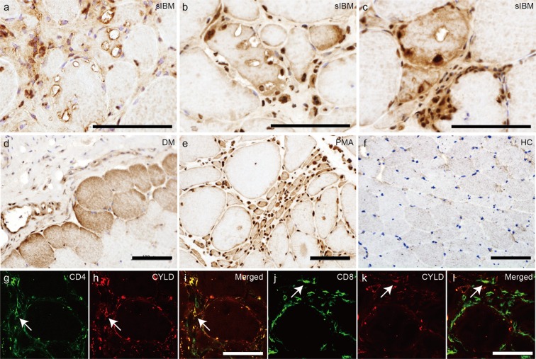
Figure 1.
Localization of CYLD in muscle fibres of sIBM patients. (a–f) Representative immunohistochemical staining for CYLD using biopsy specimens of skeletal muscles from patients with sIBM (a–c), dermatomyositis (d), progressive muscular atrophy (e), and healthy control (f). Nuclei were stained with haematoxylin. Scale bars = 100 μm. (g–l) Confocal microscopic analysis of localisation of CD4 (g) or CD8 (j) and CYLD (h,k) in muscles of patients with sIBM. Merged images are presented in (i) and (l). Arrows indicate colocalization of CD4 or CD8 with CYLD. Scale bars = 50 μm.

