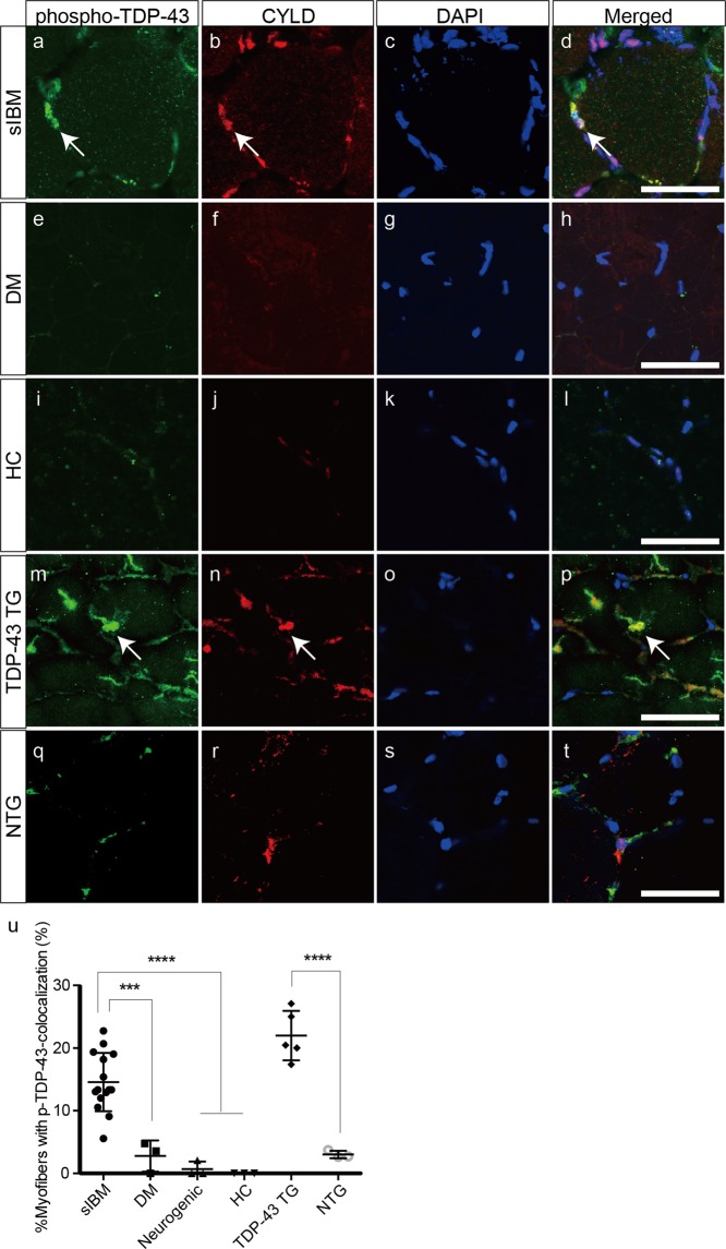
Figure 3.
Colocalization of CYLD with phosphorylated TDP-43 in muscle fibres of sIBM patients and TDP-43 TG mice. (a–t) Confocal microscopic analysis of localization of phosphorylated TDP-43 (a,e,i,m,q) and CYLD (b,f,j,n,r) in muscles of patients with sIBM (a–d), dermatomyositis (e–h), healthy control (i–l), and TDP-43 TG (m–p) or NTG mice (q–t). Nuclei were stained with 4′,6-diamidino-2-phenylindole (DAPI). Merged images are presented in (d,h,l,p,t). Arrows indicate colocalization of phosphorylated TDP-43 with CYLD. Scale bars = 50 μm. (U) Percentages of myofibres with colocalization of phosphorylated TDP-43 and CYLD in each group. n = 3–15 per group. ***P < 0.001; ****P < 0.0001.

