
Figure 5.
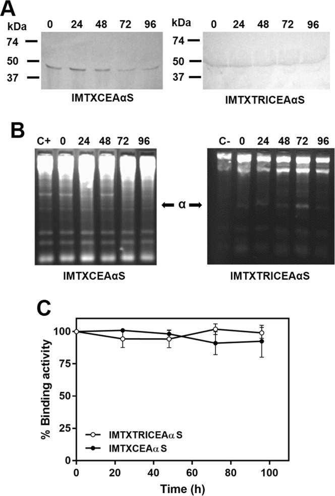
Immunotoxin stability at physiological conditions. Different aliquots of IMTXCEAαS and IMTXTRICEAαS were incubated with FBS at 37 °C for up to 96 hours. Aliquots were taken every 24 h and analyzed. (A) Western blot analysis using an anti-α-sarcin antisera. Cropped-blots are displayed. Full-length blots are presented in Supplementary Fig. S5A. Blot images were acquired and analyzed using the ChemiDoc-It (UVP) and VisionWorks LS (B) Ribonucleolytic activity assays. IMTXCEAαS gel displayed was cropped and reorganized from the original gel. Gel image was acquired and analyzed using the Gel Doc XR Imaging System and Quantity One 1-D analysis software (BioRad). The original full-length gel is presented in Supplementary Fig. S3B. (C) Flow cytometry study of immunotoxins binding to SW1222 cells.