Figure 1.
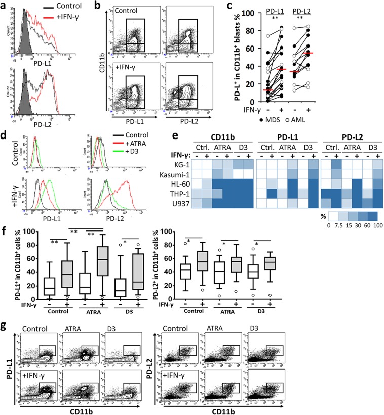
CD11b+ MDS and AML cells have increased capacity to express PD-1 ligands in response to IFN-γ. Patient-derived myeloid blasts or myeloid leukemia cell lines were stimulated with IFN-γ for 48 h. The expression of PD-L1 and PD-L2 was assessed by flow cytometry (a) without or (b) with CD11b gating. Representative plots are given. (c) Distribution of MDS and AML patients according to the change in the percentage of CD11b+ blasts with PD-1 ligand expression following the treatment with IFN-γ. (d) Representative flow cytometry histograms of PD-1 ligands are shown for HL-60 cell line pretreated with ATRA or D3. (e) Expression levels of CD11b, PD-L1, and PD-L2 following the treatment with ATRA or D3 and/or IFN-γ were schematically shown as a heat-map from the panel of myeloid leukemia cell lines screened (also refer to Supplementary Fig. 3A,D) (f) The myeloid blasts from AML or MDS patients’ bone marrow aspirates were pretreated with ATRA or D3; then, the percentage of IFN-γ-induced PD-L1 or PD-L2 expression was determined. Outliers are shown as empty circles. (g) Flow cytometry counter-plots for PD-1 ligands and CD11b are shown from a representative patient sample which was pretreated with ATRA or D3 prior to IFN-γ stimulation. (*P < 0.05, **P < 0.01; for cell lines, n ≥ 3; patient samples, n = 30).

