Figure 1.
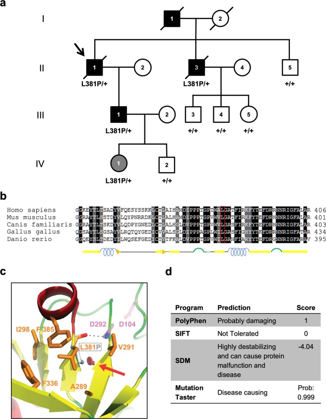
Identification of L381P renin variant in an ADTKD family. (a) Pedigree chart of the ADTKD family segregating the renin p.L381P variant. Index case (arrow): II-1. Black symbols denote clinically affected individuals, open symbols denote clinically unaffected individuals. Corresponding REN genotype is provided for each tested individual. (b) Alignment of renin sequence from human, mouse, dog, chicken and zebrafish performed with the Sequence Manipulation Suite38. Identical or similar residues are shown with a black and grey background respectively. Predicted secondary structure is shown below. (c) Detail of the human renin structure, shown in cartoon representation and colored by secondary structure (red: α-helices; yellow: β-strands; green: loops). L381 and nearby conserved hydrophobic residues (orange), as well as the two catalytic aspartates (magenta), are depicted as sticks; a red arrow indicates the steric clash (red discs) that would be introduced upon mutation of L381 to proline (grey stick). The main-chain hydrogen bond between L381 and D292 is represented by a dashed black line. The figure was made with PyMOL (Schrödinger LLC). (d) Predicted effect of the renin L381P variant, as assessed by using PolyPhen-229, Sorting Intolerant From Tolerant (SIFT)30, Site Directed Mutator (SDM)31 and Mutation Taster32.

