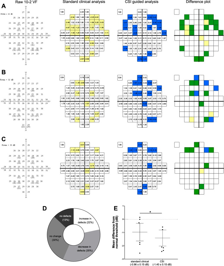
Figure 6.
Analysis of central visual function of AMD eyes using standard clinical pointwise analysis and CSI guided analysis. Examples from eyes of a (A) 73 year-old Caucasian female, (B) 72 year-old Caucasian male and (C) 77 year-old Caucasian male with intermediate AMD tested using the standard GIII stimulus and analysed in a pointwise fashion using normative distribution limits of individual test locations for GIII (standard clinical pointwise analysis) or normative distribution limits of CSIs for GIII (CSI guided analysis). Difference plots indicate defects missed by CSI guided analysis (yellow), new defects identified by CSI guided analysis (blue) or defects flagged by both analyses (green). Comparison between standard clinical pointwise analysis and CSI guided analysis for all AMD eyes (n = 23) of the number of flagged locations (i.e. defects) (D) and mean difference (± standard deviation) from sensitivity of normal eyes (E). *=p < 0.05.

