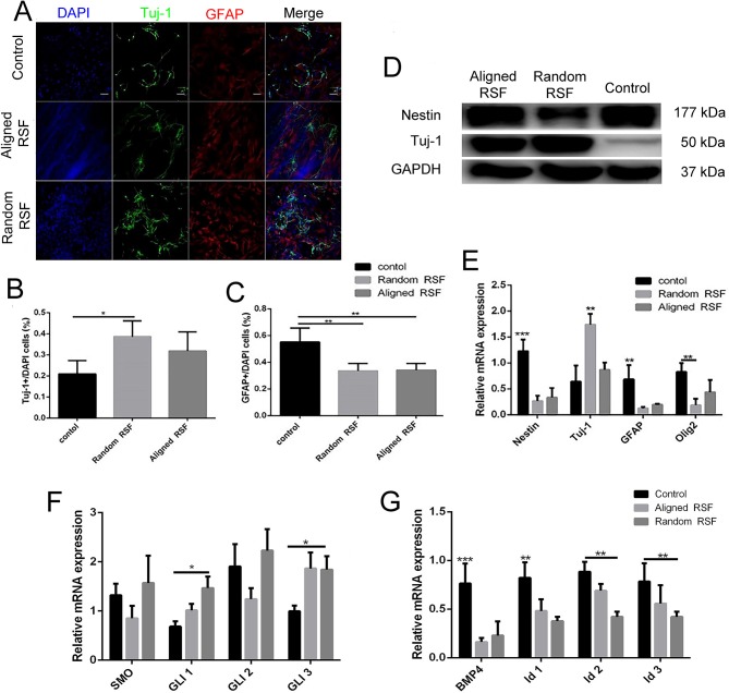
Figure 6.
The differentiation of NPCs on coverslip controls and random and aligned RSF mats after culturing for 7 days. (A) Representative fluorescence images of differentiated NPCs under differentiation conditions. The cells were immunostained with Tuj-1 for neurons (green), GFAP for astrocytes (red), and DAPI for nuclei (blue). The percentages of Tuj-1+ cells (B) and GFAP+ cells (C) among differentiated NPCs in the three groups. (D) Western blot analysis of nestin and Tuj-1 protein expression of differentiated NPCs on coverslip controls and aligned and random RSF mats. (E) qPCR analysis of nestin, Tuj-1, GFAP, and Olig1 mRNA expression in differentiated NPCs cultured on the three substrates. (F) qPCR analysis of the relative mRNA expression of Smo and Gli1–3, which are involved in the SHH signal pathway, after 7 days of differentiation on the three substrates. (G) qPCR analysis of the relative mRNA expression of BMP4 and Id1–3, which are involved in the BMP4 signaling pathway, after 7 days of differentiation on the three substrates. The data are presented as the mean ± standard error of the mean, *p < 0.05, **p < 0.01, ***p < 0.001. Scale bar: 50 μm.

