
Figure 4.
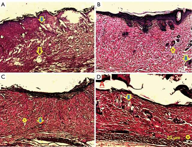
Histopathological examination of day 7 post-burned wound in control group (A), SVF group (B), PRP group (C) and SVF + PRP group (D). Yellow arrow indicates blood vessel and yellow-blue arrow indicates skin appendages (hair follicle and sebaceous gland). Hematoxylin-eosin (H&E) stained image were taken by light microscopy at 10× magnification.