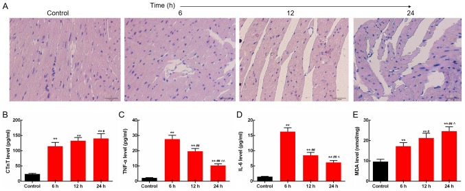
Figure 1.
LPS induces changes in myocardial morphology and inflammatory responses in rats. (A) In the septic rats, myocardial fiber rupture, interstitial edema, and inflammatory cell infiltration were observed under light microscope. Scale bar, 20 µm. (B) Serum CTnT levels were increased time-dependently. (C) TNF-α levels in the heart were increased by LPS. (D) Protein expression level of IL-6 in the heart was increased by LPS. (E) Levels of lipid peroxidation product MDA in the heart were increased time-dependently. **P<0.01 vs. the control group. #P<0.05 and ##P<0.01 vs. the 6 h group, ^P<0.05 and ^^P<0.01 vs. the 12 h group. LPS, lipopolysaccharide; CTnT, cardiac troponin T; TNF-α, tumor necrosis factor-α; IL-6, levels of interleukin 6; MDA, malondialdehyde; AMPK, AMP-activated protein kinase.

