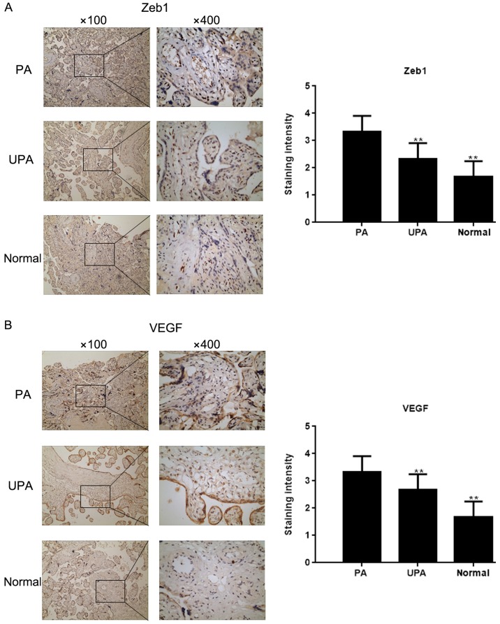
Figure 1.
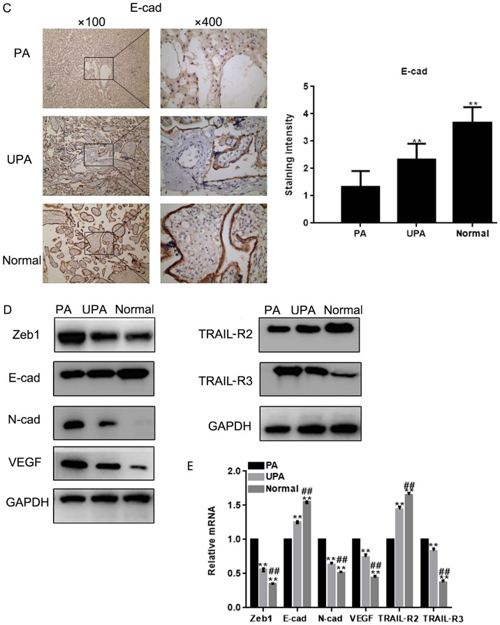
Zeb1 expression in placenta tissues. Immunohistochemical staining for the expression of (A) Zeb1 and (B) VEGF in placenta tissues (magnification, ×100 and ×400). **P<0.05 vs. PA tissues. E-cad, E-cadherin; N-cad, N-cadherin; PA, placenta accrete; TRAIL-R, TNF receptor superfamily member; UPA, placenta previa without PA; VEGF, vascular endothelial growth factor; Zeb1, zinc finger E-box-binding homeobox 1. Zeb1 expression in placenta tissues. Immunohistochemical staining for the expression of (C) E-cad in placenta tissues (magnification, ×100 and ×400). **P<0.05 vs. PA tissues. (D and E) Expression of Zeb1, E-cad, N-cad, VEGF, TRAIL-R2 and TRAIL-R3 in placenta tissues was detected by western blot analysis and reverse transcription-quantitative PCR **P<0.05 vs. PA tissues; ##P<0.05 vs. UPA tissues. E-cad, E-cadherin; N-cad, N-cadherin; PA, placenta accrete; TRAIL-R, TNF receptor superfamily member; UPA, placenta previa without PA; VEGF, vascular endothelial growth factor; Zeb1, zinc finger E-box-binding homeobox 1.


