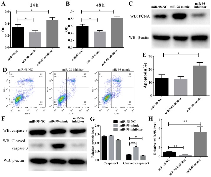
Figure 2.
Effect of miR-98 on PASMCs proliferation and apoptosis. Following transfection with miR-98 mimics or inhibitors, the hypoxia-induced PASMC proliferation for (A) 24 or (B) 48 h was analyzed by Cell Counting Kit-8 assay. (C) The protein level of PCNA was determined by western blot analysis following PASMC transfection with miR-98 mimics or inhibitors and subsequent exposure to hypoxia for 24 hz (D) Representative FACS analysis of Annexin V and PI staining. (E) Percentage of apoptotic cells analyzed by FACS. (F) The protein level of cleaved caspase 3 was determined by western blot analysis following PASMC transfection with miR-98 mimics or inhibitors and subsequent exposure to hypoxia for 48 h. (G) Densitometry analysis of the western blots of caspase 3 and cleaved caspase 3. (H) The miR-98 expression in PASMCs transfected with miR-98 mimics or inhibitors was examined by reverse transcription quantitative polymerase chain reaction. *P<0.05 and **P<0.01 (n=3). miR, microRNA; PASMCs, pulmonary artery smooth muscle cells; PCNA, proliferating cell nuclear antigen; PI, propidium iodide; FACS, fluorescence-activated cell sorting; NC, negative control.

