Figure 4.
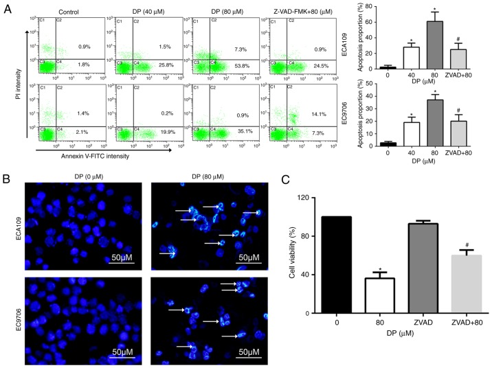
DP induces caspase-dependent apoptosis in ESCC cells. (A) Apoptosis was analyzed by flow cytometry using Annexin V-FITC/PI double staining assay. DP (0, 40 and 80 µM) treatment for 24 h significantly increased the proportion of apoptotic cells that was reversed by pretreatment with Z-VAD-FMK for 1 h in ECA109 and EC9706 cells. (B) Morphological changes of apoptosis were observed under fluorescence microscope using Hoechst 33342 staining. After DP (0 and 80 µM) treatment for 24 h, chromatin condensation and DNA fragmentation (as indicated by arrows), were observed in ECA109 and EC9706 cells. (C) After pretreatment with Z-VAD-FMK (50 µM), reduction in cell viability mediated by DP treatment was partly reversed in ECA109 cells. The data are expressed as the mean ± standard deviation (n=3). *P<0.05 vs. control; #P<0.05 vs. DP treatment. DP, dracorhodin perchlorate; ESCC, esophageal squamous cell carcinoma. ZVAD+80, cells pretreated with Z-VAD-FMK and then treated with 80 µM DP.

