Figure 1.
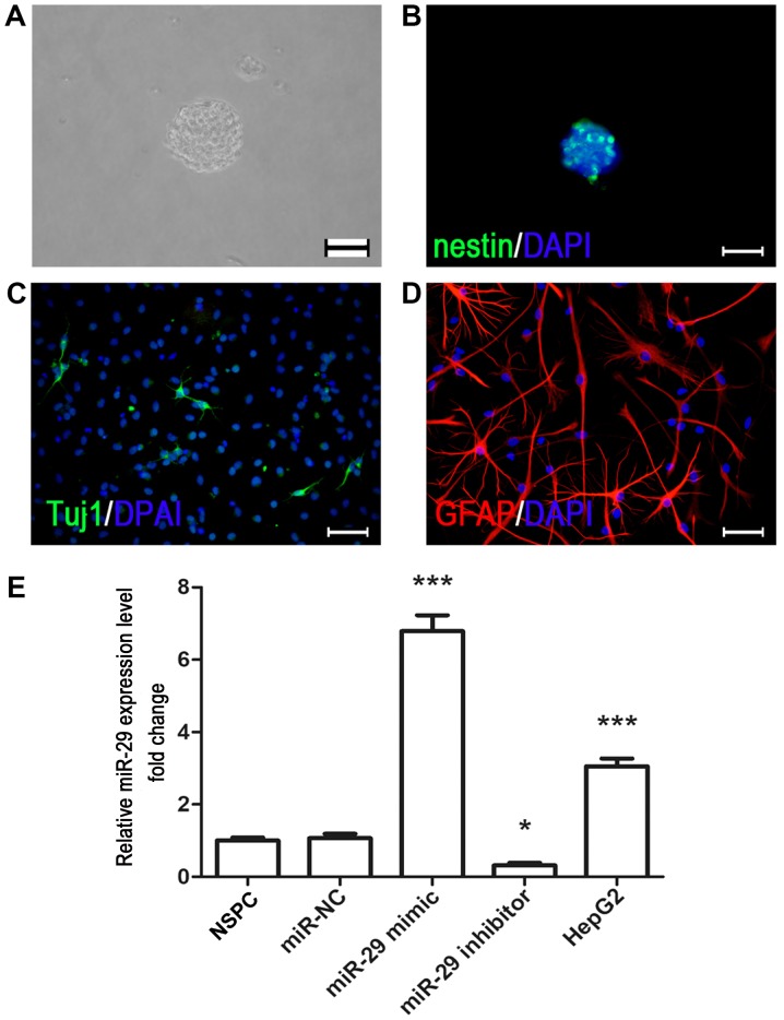
Culture and immunocytochemical characterization of fetal rat cortex NSPCs. (A) NSPCs were isolated from the E14.5 fetal rat cortex, and neurospheres were formed following 3–5 days of cell proliferation. Scale bar, 200 µm. (B) Nestin (green) immunostaining of NSPCs was conducted in neurospheres under proliferating conditions. Scale bar, 200 µm. NSPCs were differentiated into (C) Tuj1- and (D) GFAP-immunopositive cells by removal mitogens and adding 1% fetal bovine serum. Scale bar, 40 µm. Nuclei were counterstained with DAPI (blue). (E) Reverse transcription-quantitative PCR analysis indicated that miR-29 mimic increased the expression of miR-29, whereas miR-29 inhibitor downregulated its expression. The liver cancer cell line HepG2 was used as a positive control. *P<0.05, ***P<0.001 vs. miR-NC. NSPC, neural stem/progenitor cell; miR-29, microRNA-29; miR-NC, microRNA negative control; Tuj1, neuron-specific class III β-tubulin; GFAP, glial fibrillary acidic protein.

