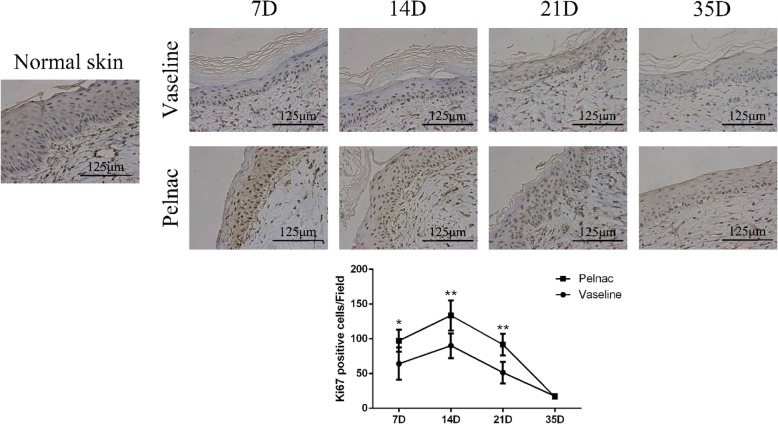
Fig. 9.
Ki-67 expression in wound tissue of rats at different time points post-operation after treatment with Vaseline gauze or Pelnac as overlay. (Top) Ki-67 immunohistochemistry was performed on days 7, 14, 21, and 35 (7D, 14D, 21D, and 35D, respectively) (× 400). Scale bars, 125 μm. (Bottom) quantitative analysis of Ki67-positive cells. Data are presented as mean ± standard devistion. Error bars indicate standard deviation. Statistical analysis was performed by repeated-measures ANOVA. *p < 0.05, **p < 0.01

