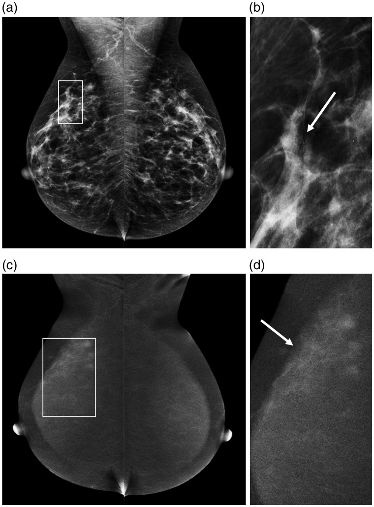
Fig. 4.
Example of a 52-year old patient recalled for suspicious breast calcifications in the right breast. (a) The LE images with a detailed view of the calcifications in (b) (arrow). After contrast administration, the recombined images (c) show an area of segmental non-mass enhancement, showing that the true disease extent is much larger than was initially suspected on conventional mammography (d, arrow). Final histopathological results revealed a small invasive ductal carcinoma (6 mm) surrounded by grade 2 DCIS (40 mm).

