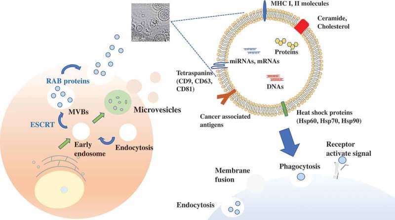
Figure 1.

EV production procedure.
Exosomes, 50–150 nm, are initially formed by a process of inward budding in early endosomes to form multivesicular bodies. Microvesicles are larger than exosomes, approximately 100–1000 nm. They are composed of lipid components and are directly shed or budded from plasma membranes. RAB proteins (RAB27A, RAB27B), ESCRT (Alix, TSG101) are associated with EV secretion. There are some markers on EV membranes, which is useful to detect EVs. EVs vary in size, properties, and secretion pathway depending on the originated cells, and the EVs are indeed taken up by recipient cells via a variety of mechanisms. Electron microscopy indicates the EVs derived from MCF7 breast cancer cell lines.
