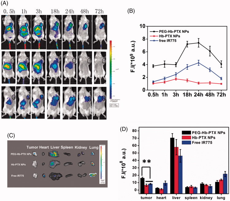
Figure 4.
In vivo fluorescence imaging and biodistribution of free IR775, Hb-IR775, and PEG-Hb-IR775 NPs in H22 tumor-bearing mice. (A) NIR fluorescence imaging after intravenous injection of PEG-Hb-IR775 NPs, Hb-IR775 NPs, and free IR775 (tumors indicated by black circle). The colors indicated that the fluorescence intensity in the picture increased from blue to red. (B) Fluorescence intensity (F.I) of IR775 in the tumors of three groups. (C) Ex vivo fluorescence images of tumors and major organs after administration at 72 h. (D) Semiquantitative assessment of biodistribution of IR775 after injection. **p < 0.01.

