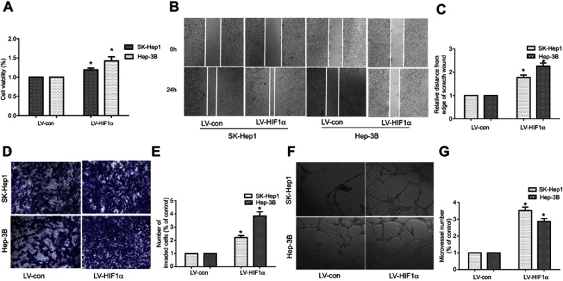
Figure 4.
HIF1α overexpression promotes the proliferation, migration, invasion, and angiogenic ability of SK-Hep1 and Hep-3B cells.
Notes: (A) Cell proliferation was assessed by the CCK-8 assay. (B) Cell migration was assessed by the wound healing test. (C) The histogram represents the migration distance relative to the control 24 h after scratching the cell layer. (D) Cell invasion was examined by the transwell invasion test. (E) The histogram represents the average number of invasive cells relative to the control. (F) HCC cell-induced angiogenesis of HUVECs was studied by the tube formation test. (G) The representative histogram of the area covered by the tube network was quantified using the Image-Pro Plus software. Data were represented with the GraphPad Prism 5.02 software. *P<0.05, compared to the control group.
Abbreviations: HCC, hepatocellular carcinoma; HUVECs, Human umbilical vein endothelial cells.

