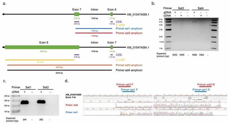
Figure 8.

Detection of 3ʹUTR sequences of YAP1 transcripts in bovine muscle. (a) Design of primer sets for YAP1 transcript XM_015474584.1 and XM_015474589.1. Black arrows = YAP1 gene. Green rectangles = exons. Purple lines = coding sequences. Yellow lines = 3ʹUTR sequences. Blue/red/brown/black lines = amplicon sequences of different primer sets. Numbers indicate expected length of each sequence. (b) PCR products of primer set3 and set4 with genomic DNA (gDNA), cDNA, and water (-/-) as templates on 0.5% agarose gel. (c) PCR products of primer set1 and set2 with cDNA and water (-) as templates on 1% agarose gel. (d) Sanger sequencing results of the two bands detected in (c). Locations of the primers are indicated on the top. F = forward primer. R = reverse primer.
