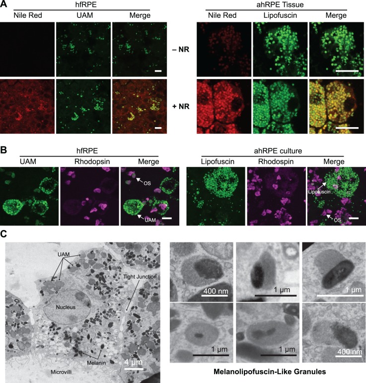
Figure 2.
Composition of UAM. (A) Nile Red staining of neutral lipids associated with UAM in hfRPE (left) and in vivo lipofuscin in freshly isolated RPE tissue (ahRPE) from 69-year-old male with no ocular pathology (right). Top row is without Nile Red to control for any autofluorescence bleedthrough from UAM or lipofuscin into Nile Red channel. Bottom row is with Nile Red. Scale bar: 10 μm. (B) Rhodopsin staining is not associated with hfRPE UAM (left) or in vivo lipofuscin “carried-over” into P1 ahRPE primary cultures (right). Cultures were fed a single bolus of OS as a positive control to confirm rhodopsin staining and demonstrate intrinsic autofluorescence from OS. UAM/lipofuscin and OS autofluorescence (green), B630 anti-rhodopsin antibody (purple). Scale bar: 10 μm. (C) (Left) Electron micrograph of hfRPE fed photo-oxidized OS on 20 separate days over a 4-week period reveals a homogenous granular structure to the accumulated UAM, similar to in vivo lipofuscin. (Right) Higher magnification views from other cells show that certain granules contain a mixture of UAM and melanin, mimicking the appearance of melanolipofuscin in RPE from patients as they age.

