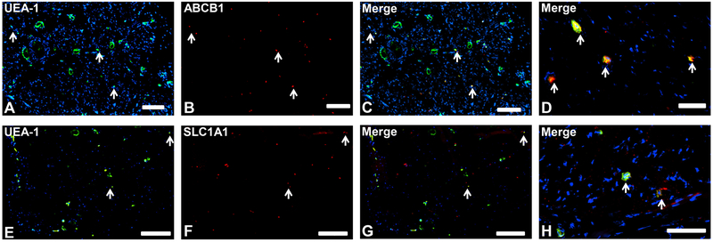
Figure 1. Blood-nerve barrier-restricted proteins.
Representative indirect fluorescent digital photomicrographs of cryostat axial sections of normal adult sural nerves show UEA-1-positive endothelial cells (green; A and E) with proteins ABCB1 and SLC1A1 (red; B and F respectively) demonstrating restricted expression by endoneurial microvascular endothelium on merged images (yellow; C and G respectively), further demonstrated at higher magnification (yellow/orange; D and H respectively). This suggests that these proteins have restricted BNB functions. White arrows demonstrate positively staining endoneurial microvessels. Blue (DAPI) staining indicates nuclei. Scale bar 500 μm for A-C and E-G, and 125 μm for D and H.

