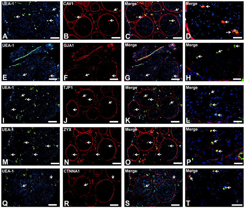
Figure 4. Endothelial and specialized epithelial-specific proteins.
Representative indirect fluorescent digital photomicrographs of cryostat axial sections of normal adult sural nerves show UEA-1-positive endothelial cells (green; A, E, I, M, Q) with expression of CAV1, GJA1, TJP1, ZYX and CTNNA1 (red; B, F, J, N, R respectively) restricted to endoneurial microvessels, fenestrated epineurial macrovessels and the perineurium, shown in the merged images at lower and higher magnifications (yellow/ orange for endothelial cells). Expression of these proteins by both microvascular and macrovascular endothelial cells and the perineurium suggests non-restrictive barrier, but specialized endothelial and epithelial cell functions in the normal adult human peripheral nerves. White arrows demonstrate positively staining endoneurial microvessels. Blue (DAPI) staining indicates nuclei. Scale bar 500 μm for A-C, E-G, I-K, M-O, and Q-S and 125 μm for D, H, L, P and T.

