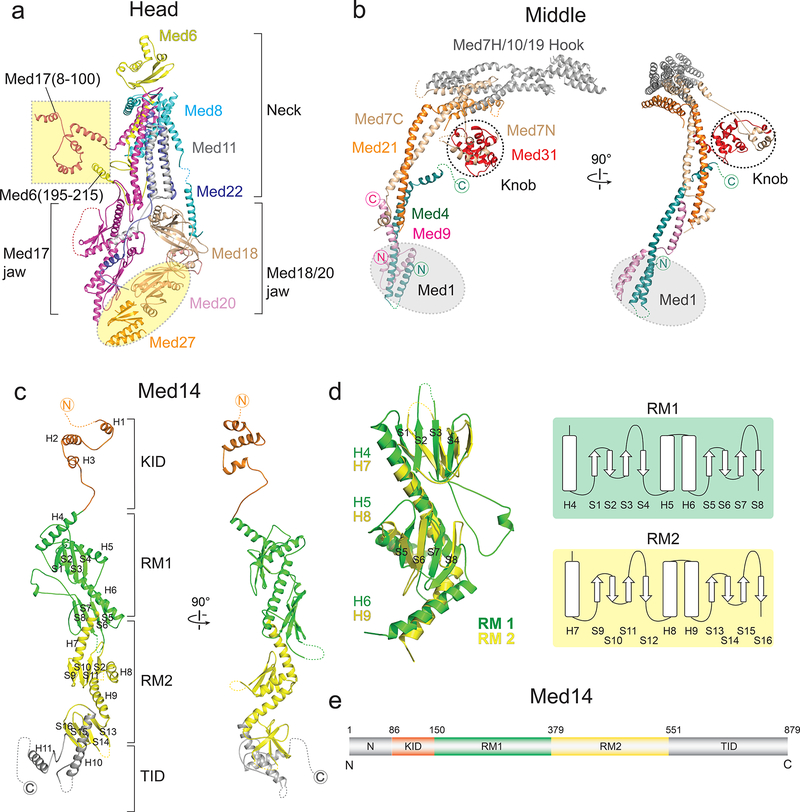
Figure 2. Structures of the Head, Middle, and Med14.
a, Head module. Med20, Med27, Med17(8–100) and Med6(195–215), absent in a previous X-ray structure, are highlighted in yellow. b, Middle module. Non-Middle subunits in the knob (indicated by dashed circle) are not shown. Med1 position highlighted in gray. c, Med14 (81–686) includes two repeats of a structural domain (RM1 and RM2; in green and yellow), a C-terminal Tail interaction domain (TID; in gray), and an extended N-terminal knob interaction domain (KID; in light orange) that forms part of the Middle’s knob. Alpha helices and beta sheets are labeled. d, Overlay of Med14’s RM1 and RM2 and corresponding topology diagrams. Alpha helices and beta sheets labeled as in (c). RMSD values between 136 atom pairs in the RM1 and RM2 portions of Med14 is 5.4 Å. e, Med14 domain architecture.

