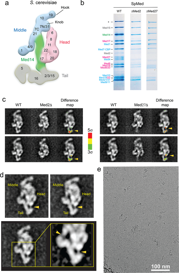
Extended Data Figure 1. S cerevisiae and S pombe Mediator subunit localization.
a, Localization of S cerevisiae Mediator subunits. b. Coomassie blue-stained SDS-PAGE analysis (4–20% gradient gel) of purified wild-type, ΔMed2, and ΔMed27 S pombe Mediator. The Med2 band is comparatively weaker, suggesting that the subunit might be substoichiometric in purified wild-type SpMED. Deletion of Med2 results in loss of Med27, but not the other way around. The band marked with the asterisk corresponds to Rpb1, which was confirmed by mass spectrometry. Subunits are colored by module (Head, pink; Middle, blue; Tail, gray) c. 2D class averages for wild-type, ΔMed2, and ΔMed27 SpMED, and color-coded (by standard deviation values from the average) difference maps indicating the position of Med2 and Med27 (highlighted by yellow arrow heads). d, Wild-type SpMED class averages and a close up showing a Med27-Tail connection (lower-right arrowhead) comparable to the connection between the Med4/Med9 4-helix bundle and the rest of the Middle module (upper-left arrowhead). e, A raw micrograph showing a typical area of a SpMED cryo-EM sample.

