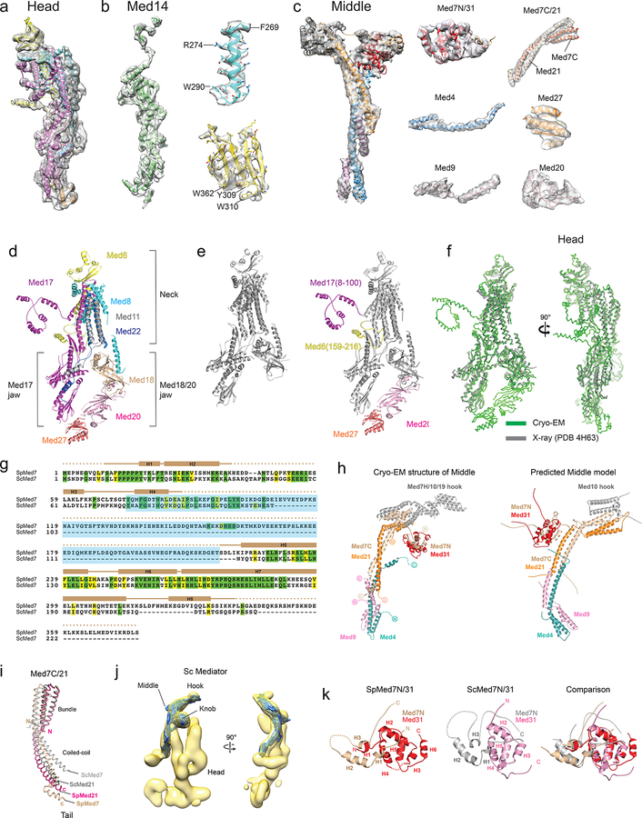
Extended Data Figure 3. Cryo-EM map and structure of SpMED Head and Middle modules.
a, Head portion of the SpMED cryo-EM map showing secondary structure elements corresponding to individual subunits. The resolution of the Head portion of the map 4.5–5.0 Å. b, Med14 portion of the EM map and close-ups showing density corresponding to bulky Med14 side chains. The resolution of the Med14 portion of the map 4.0–4.5 Å. c, Middle portion of the SpMED cryo-EM map showing secondary structure elements corresponding to various subunits, and map segments corresponding to individual Middle components. The resolution of the Middle portion of the map and its segments is 5–6 Å. d, Cryo-EM structure of the Head module. Colors as indicated in Fig 1c. e, Partial X-ray structure of the Head module (PDB 4H63; left, in gray) compared with the cryo-EM structure of the Head module (right) with portions not included in the X-ray crystal structure colored and labeled. f, Superposition of the X-ray (gray) and cryo-EM (green) structures of the Head module shows a close correspondence between common elements, indicating that the overall conformation of the Head is not changed by interaction with other Mediator modules. RMSD values between 744 Cα atom pairs in corresponding portions of the X-ray and EM structures of the Head is 2.1 Å. g, Alignment of SpMed7 and ScMed7 protein sequences. The secondary structure evident in the cryo-EM structure of SpMed7 is indicated. Identical and similar residues are highlighted in green and yellow, respectively. Residues in S pombe Med7 expected to be part of the hook are highlighted in light blue. h, Cryo-EM structure of the Middle (left) and a model of the Middle41 based on X-ray structures of Med7C/Med21 and Med7N/31, homology modeling of Med4/9/10, and results from cross-linking mass spec analysis (right). i, Comparison between the X-ray structure of Sc Med7N/21 (gray) and the Sp Med7N/21 from the cryo-EM map. j, Fitting of the Sp Middle module structure (solid blue) into the Sc Mediator cryo-EM map (transparent yellow) shows that the Middle structure is conserved between S cerevisiae and S pombe. k, Comparison between the cryo-EM structure of SpMed7N/31 and the X-ray structure of ScMed7N/31 shows that this portion of the Mediator structure is highly conserved. RMSD values between Cα 84 atom pairs in corresponding portions of the X-ray structure of ScMed 7N/31 (PDB 3FBI) and the EM structure of SpMed 7N/31 is 2.0 Å.

