
Figure 12:
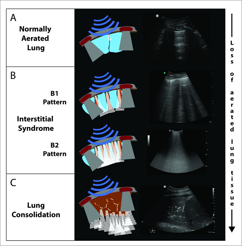
Schematic and still images of lung ultrasound in normal lung (Panel A), interstitial syndrome (Panel B) and alveolar syndrome (Panel C). In the normally aerated lung (Panel A), the findings include a homogenous pleural line (uppermost horizontal white line in image), the presence of an A line (i.e. short horizontal white line in mid-image, an artefact from the pleural line), lung sliding (see respiratory changes seen in the dynamic video- Supplemental Digital Content 6), and a lung pulse (see cardiac changes seen in the dynamic video- Supplemental Digital Content 6). The interstitial syndrome (Panel B) involves loss of lung aeration and is of two types. The ‘B1’ pattern, corresponding to moderate loss of aeration, has 3 or more B-lines (vertical) per intercostal space, whereas the ‘B2’ pattern, corresponding to more severe loss of aeration, has multiple coalescent B-lines per intercostal space. Lung consolidation (Panel C), indicates substantially increased density with almost complete loss of aeration. This is characterized by an anechoic (i.e. tissue-like) image arising from the pleural line.