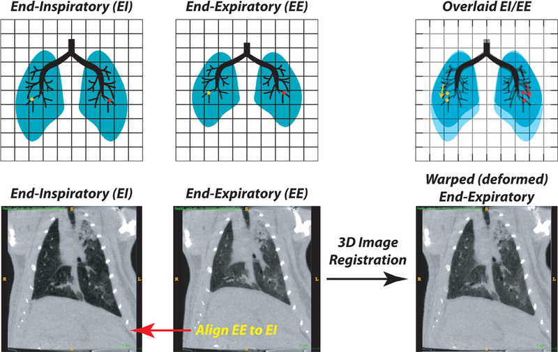
Figure 3:

Schematic showing the process of image registration between end-inspiratory (EI) and end-expiratory (EE) images (Upper Panels - Schematic; Lower Panels - representative CT slices). The end-expiratory image is expanded in three dimensions to align all visible tissue features, including airways and blood vessels, to the target end-inspiratory image. With registration of tissue points, it is then possible to track the displacement that any point in the image undergoes during each tidal deflation (e.g., the red and yellow dots). The product of the registration is thus a ‘warped’ (i.e. constrained fit) end-expiratory image. An example of image registration performed on end-inspiratory and end-expiratory CT scans obtained in a ventilated rat after lung injury is shown (Bottom Panel; see also Supplemental Digital content 1).
