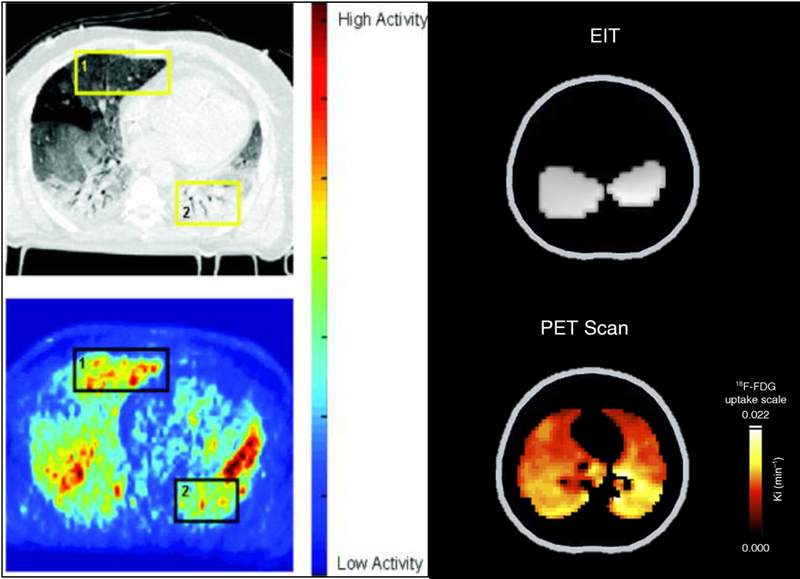
Figure 8:

Paired Computed tomography (CT, Upper Left Panel) and [18F]-fluoro-2-deoxy-d-glucose (18FDG) positron emission tomography (PET, Lower Left Panel) from a patient with ARDS. A high level of 18FDG activity is seen in the in ventral lung in the PET scan (Yellow Box 1) that appears normally aerated in the CT scan (‘baby lung; Black Box 1). Paired electrical impedance tomography (EIT, Upper Right Panel) and PET (Lower Right Panel) images are shown from a pig with lung injury ventilated with low tidal volume and low PEEP, while performing strong inspiratory effort. The EIT image shows regions of maximum ventilation (grey shade) in dependent lung near the diaphragm, and the PET image shows high FDG activity, indicating inflammation, in the same dependent regions. Reproduced with permission from Refs. 168 and 170.
