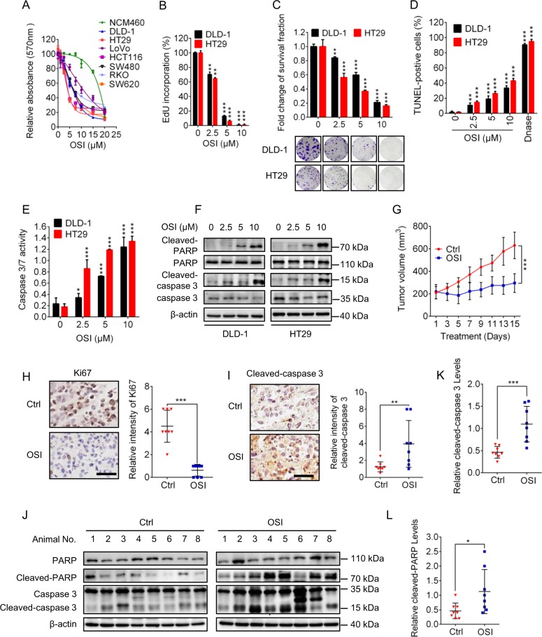
Fig. 1. OSI inhibits the growth of CRC cells in vitro and in vivo.
a The relative absorbance at 570 nm of CRC cell lines (DLD-1, HT29, LoVo, HCT116, SW480, RKO) and noncancerous colorectal cell line NCM460 treated with indicated concentrations of OSI for 24 h. b, c CRC cells were treated with indicated concentrations of OSI for 24 h. Cell proliferation was detected by EdU incorporation assay (b) and colony formation assay (c). d–f In situ TUNEL assay (d), caspase 3/7 activity assay (e), cleaved-PARP and cleaved-caspase 3 detection (f) were used to examine the apoptotic effects in OSI-treated CRC cells. g The volume of tumors of mice in cohorts treated with vehicle (n = 8) or OSI (n = 8) was measured at the indicated time points. h The Ki67 expression of tumors was detected by IHC. Scale bar, 50 μm (Left). i The Ki67 expression of tumors was detected by IHC. Scale bar, 50 μm (Left). j Immunoblotting analysis of cleaved-caspase 3 levels in tumor xenografts obtained from vehicle- or OSI-treated mice. (Each protein of interest from each group was electrophoretically transferred onto a PVDF membrane, incubated with indicated primary and secondary antibodies, and developed as a digital image.) k Quantification of cleaved-caspase 3 level in (j). l Quantification of cleaved-PARP level in (j) **P < 0.01; ***P < 0.001

